
Non-viral Gene Editing of Marrow Stem Cells Shows Potential in Early Study


A non-viral, CRISPR-based gene-editing tool showed a potential to reverse mutations that cause sickle cell disease (SCD) by successfully editing genes in stem cells within the bone marrow of mice.
Data from this proof-of-concept study also found that changes in stem cell genes were sustained for more than one year after a single course of treatment.
“This new data supports the possibility of delivering a safer solution to treat blood disorders, including sickle cell disease, by avoiding the need for bone marrow transplantation,” John Leonard, MD, president and CEO of Intellia Therapeutics, the technology’s developer, said in a press release.
Study findings were presented by Sean Burns, MD, senior director of Intellia’s Disease Biology and Pharmacology group, in the presentation “In Vivo Genome Editing of Hematopoietic Stem and Progenitor Cells,” given at the Keystone eSymposium: Precision Engineering of the Genome, Epigenome and Transcriptome, held online in March.
SCD is caused by mutations in the HBB gene, which carries instructions for a subunit of hemoglobin, the protein found in red blood cells that transports oxygen throughout the bloodstream. These mutations result in sickle-shaped red blood cells that can clog small blood vessels, causing pain crises.
New targeted gene-editing technologies, such as CRISPR/Cas9, have the potential to reverse mutations in the HBB gene, restoring the production of healthy hemoglobin, and preventing red blood cell sickling.
A clinical study demonstrated that ex vivo [outside a living organism] gene editing can be beneficial for certain SCD patients. This type of gene editing involves harvesting blood cell precursors, known as human hematopoietic stem cells (HSCs), from a patient’s bone marrow. Once the HBB gene sequence is edited, blood cell progenitors are ready to be returned to the patient.
The genetically modified stem cells then settle within the patient’s bone marrow, and start giving rise to all types of blood cells, including red blood cells, effectively replacing those that are defective.
However, this approach requires patients to complete a course of treatment — usually consisting of a combination of different chemotherapy agents — to destroy the faulty blood cell progenitors, and “make room” within the bone marrow for the genetically modified stem cells.
This treatment can put patients at a higher risk of developing certain types of cancer, particularly leukemia, as well as affect their fertility.
Because these medications wipe out a patient’s entire bone marrow, immune cell production is compromised and the immune system less able to fight against disease-causing microbes. As a result, patients are more susceptible to infections. Stem cell transplants done using this approach are also reported to be limited by a patient’s comorbidities (co-existing conditions), their high cost, and need for specialized care.
Scientists at Intellia report having developed an alternative in vivo approach to editing the HBB gene in HSCs within the bone marrow itself, that may be simpler than the cell manufacturing process required for ex vivo gene editing, and without a need for as extensive post-treatment care.
In this method, CRISPR/Cas9 gene-editing components are delivered directly to target cells by lipid nanoparticles (LNPs), instead of a modified virus. LNPs are small biodegradable vesicles that are unlikely to trigger an immune reaction, and can easily be produced at a large scale.
LNP technology has been previously used to target cells in the liver. However, in this proof-of-concept study, LNPs were designed and tested in vivo — in animals — to identify specific compositions that resulted in enhanced delivery of genetic material to the animals’ bone marrow and HSCs.
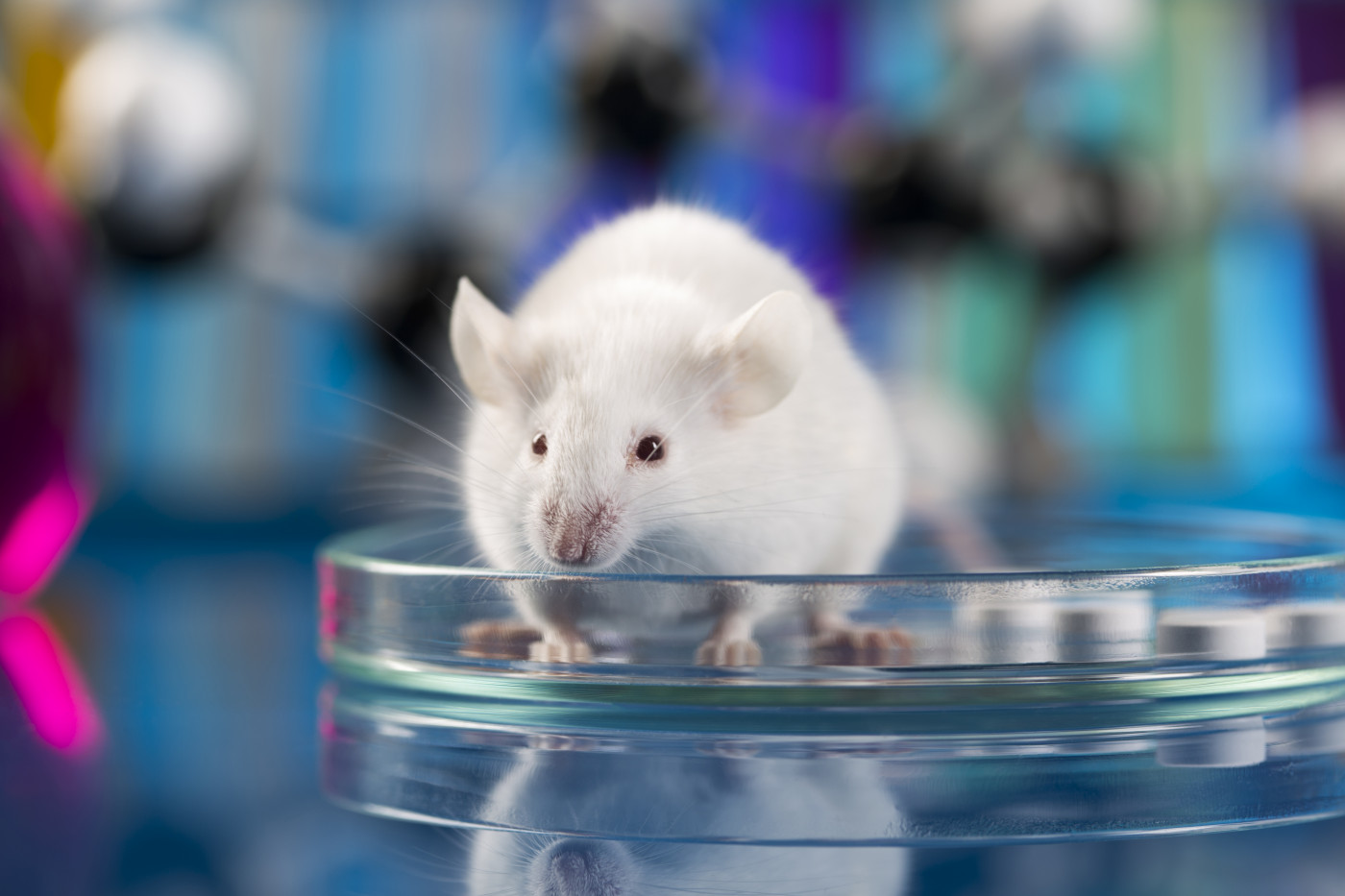
Researchers injected different doses of CRISPR/LNPs into the tail vein of healthy (wild type) mice.
Analyses performed a week later found a dose-depended gene editing in the animals’ entire bone marrow, and HSCs at levels predicted to have a therapeutic benefit in SCD patients. Editing levels were sustained for at least one year and increased with multiples successive treatment doses.
A competitive bone marrow transplant test, which is normally used to assess stem cells’ ability to give rise to different types of cells, demonstrated that genetically modified HSCs retained their ability to grow into all types of blood cells, including immune cells. At the same time, a population of edited HSCs was seen to remain stable within the animals’ bone marrow for more than 14 weeks.
Investigators then performed a series of experiments on humanized mice, or animals given a transplant of human cells containing the CD34 protein, a marker of human HSCs.
A bone marrow examination showed that a single dose of LNPs led to a clinically significant level of editing in human CD34-positive cells, suggesting “cross-species relevance of [the] LNP platform for genome engineering of [HSCs],” the team wrote.
“We’ve demonstrated we can expand our in vivo capabilities originally designed for liver applications to other tissues and achieve therapeutically meaningful levels of gene editing,” Leonard said.
The company plans to further demonstrate the approach’s clinical relevance by confirming these results in non-human primates.