
Low Oxygen Regions in Brains of Sickle Cell Patients are Prone to Small Strokes, Study Shows
Written by |
Sickle cell disease patients experience silent strokes in the deep white matter of the brain that is prone to low oxygen supply, a brain-mapping study shows.
The study, “Silent infarcts in sickle cell disease occur in the border zone region and are associated with low cerebral blood flow,” was published in the journal Blood.
Children with sickle cell anemia (SCA) are at a high risk of experiencing silent cerebral infarctions (SCIs). These events, although seemingly small, can contribute to disease progression, cause cognitive impairment, and lead to a full-blown stroke in children and adults with SCA.
Low blood supply (anemic events) and high blood pressure have been identified as risk factors for SCI; however, its mechanisms are not entirely clear. It is hypothesized that regions of the brain that experience an SCI have poor blood and thus poor oxygen supply (hypoxia), according to the study.
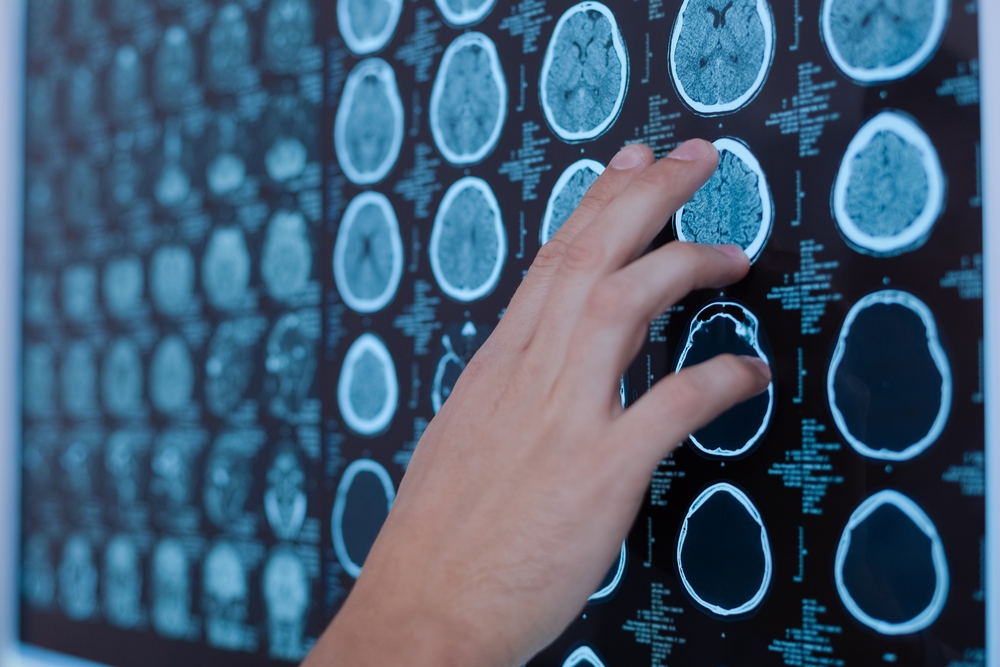
Researchers evaluated the brain density maps of 286 children (5–15 years old) with SCI who participated in a Phase 3 silent infarct transfusion (SIT) trial (NCT00072761), a multicenter international study where participants had a diagnosis of SCA or sickle cell thalassemia and had at least one brain lesion that resembled an infarct by magnetic resonance imaging (MRI).
In the SIT trial, the patients were randomly assigned to receive either the standard care for SCI or blood transfusion therapy.
The researchers first identified the SCI lesions that were imaged as part of the SIT trial in the 286 children. They then mapped each SCI to a brain atlas to create an overlay they called an “SCI density map.” This map was divided into regions of low (1%–6%), moderate (7%–12%), and high (13%–18%) infarct density.
The map showed that 90% of infarct lesions occurred in border regions of the deep white matter within the brain. These appeared as regions of high density and made up only 5.6% of the brain volume.
Next, another group of 41 children with SCA between the ages of 5 and 21 years were recruited to analyze blood flow to the brain during a period of no infarction using arterial spin labeling MRI. This resting cerebral blood flow helped the researchers determine if the blood supply is interrupted to the regions identified in the children in the SIT study.
They confirmed that areas of reduced resting blood flow overlapped the regions of high density observed in SIT children, confirming the hypothesis that infarctions occur in areas of low blood supply.
“The infarct density map provides a template for future studies evaluating novel imaging biomarkers predicting stroke risk in SCA, and will assist neuroradiologists in identifying SCIs within this established region,” the study concluded.


Leave a comment
Fill in the required fields to post. Your email address will not be published.