Important Enzyme Found Deficient in Lung Blood Vessels of Patients

An enzyme known to play a key role in blood vessel health is present at low levels in the cells lining the lung blood vessels of patients with sickle cell disease (SCD), which may affect how these cells stick to their supporting matrix, a study has found.
Lack of this enzyme, called superoxide dismutase 2 (SOD2), increases oxidative stress — a form of cellular damage resulting from an imbalance in oxidant molecules — and can lead to blood vessel disease.
Researchers also found that SOD2 deficiency prevented fibronectin — a protein that helps wounds heal — from combining with other fibronectin molecules in so-called dimers (a molecule or molecular complex consisting of two identical molecules linked together). This affected how much fibronectin was released from cells and how well cells were able to increase in number, move around, and prevent certain substances from passing through.
“These data suggest that further investigation into therapeutic treatments targeting SOD2-mediated changes … may be beneficial in SCD,” the researchers wrote.
The study, “Endothelial superoxide dismutase 2 is decreased in sickle cell disease and regulates fibronectin processing,” was published in the journal Function.
SCD is a genetic condition in which red blood cells acquire an unusual sickle-like shape that makes them more rigid and sticky.
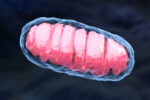
SOD2 is located in mitochondria — the tiny structures that make most of a cell’s energy. There, SOD2 speeds up the conversion of superoxide to hydrogen peroxide, two unstable molecules that contain oxygen and can easily react with other molecules in a cell. SCD is marked by an increase in oxidative stress due to high levels of such reactive oxygen species.
Moreover, a decrease in SOD2 blood levels is linked to an increased breakdown of red blood cells (hemolysis), inflammation, and heart muscle disease (cardiomyopathy) in SCD.
Now, researchers at the University of Pittsburgh and Carnegie Mellon University in Pennsylvania have discovered SOD2 is present at low levels in the cells lining the lung blood vessels of SCD patients.
First, they prepared thin slices of lung tissue obtained from two men and two women with SCD. Their mean age was 44.2 years. Lung tissue from four age- and sex-matched individuals was used as a control.
The researchers found about 40% less SOD2 was present in tissue samples from patients compared with controls. The reduction was observed both in endothelial cells — those lining the inside of blood vessels — and smooth muscle cells, which help blood vessels contract and relax.
Using a mouse model of SCD known as the Townes model, the researchers observed a decrease of about 25% in lung SOD2 levels in mice with the disease compared with those that did not have the disease. Townes model mice carry a mutated version of the human beta-globin (HBB) gene, which causes SCD.
To understand the role of SOD2 in the lining of blood vessels, the researchers used siRNA, a type of RNA that can block the production of proteins, to decrease SOD2 levels in human endothelial cells obtained from tiny blood vessels in the lungs.
A decrease in SOD2 levels resulted in a marked increase in superoxide levels in mitochondria, which became damaged, but did not lose their capacity to produce energy.
Next, the researchers watched for changes in the way endothelial cells behaved. They found that layers of endothelial cells with low SOD2 levels were leakier, grew less, and were less able to migrate than those with normal SOD2 levels.
“SOD2 is essential in the maintenance of multiple endothelial cell functions,” the researchers wrote.
They also found that low SOD2 levels resulted in a decrease in the production of hydrogen peroxide in mitochondria. In turn, this led to reduced assembly of fibronectin molecules into dimers, “an essential step in the formation of fibronectin fibrils and integration into the extracellular matrix.”
“These results demonstrate that endothelial cells are deficient in SOD2 expression in SCD patients and suggest a novel pathway for SOD2 in regulating fibronectin processing,” the researchers wrote.


