Brain development changes seen in children with sickle cell anemia
Study shows subtle brain differences in children without prior stroke or infarction
Written by |
Children with sickle cell anemia (SCA), the most common and severe form of sickle cell disease (SCD), show signs of altered development in specific regions of the brain, even in children without a history of silent or overt stroke, a new study from the U.S. reports.
Using an artificial intelligence (AI) tool that estimates a person’s “brain age” based on MRI scans, researchers found that overall brain development appeared similar to that of healthy children, but certain brain regions in those with SCA appeared “younger” than expected for their age — a sign of altered brain development.

Regional brain changes seen even without stroke or infarction
These changes were observed in children without a history of silent or overt stroke and were found in regions that overlap with areas of the brain at higher risk of infarction, or tissue death due to inadequate blood supply. This suggests that early, subtle changes in brain development may occur in SCA even without a history of infarction.
“We conclude that there is a regional decrease in brain age in children with SCA without stroke compared with controls, suggesting altered brain development,” the researchers wrote, noting that “solely focusing on infarct status alone ignores earlier stages of neurological injury in this vulnerable population.”
The study, “Regional brain age is decreased in children with sickle cell anemia,” was published in Blood Advances.
SCA is caused by mutations that lead to the production of an abnormal form of hemoglobin, the protein in red blood cells that carries oxygen. This defective protein causes red blood cells to become rigid and sickle-shaped, making them more likely to break down and block small blood vessels.
The breakdown of red blood cells results in anemia and can trigger inflammation throughout the body. At the same time, their tendency to block small blood vessels impairs blood flow and reduces oxygen delivery to tissues.
In the brain, this blood vessel blockage can prevent some regions from receiving enough oxygen and nutrients, causing damage and infarction, which may be silent or occur as part of an overt stroke. This can also contribute to cognitive problems, independently of strokes.
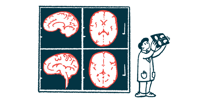
MRI studies have long explored brain effects in sickle cell
Researchers have long used brain MRI scans to study how SCA affects the developing brain, with previous studies suggesting changes in brain volume, structure, and connectivity. However, many of these approaches rely on specialized imaging techniques and complex analyses that limit their use in routine clinical care.
DeepBrainNet is an AI tool that has been used in adults with SCA to estimate brain age based on a single brain MRI scan. It was trained on large sets of MRI scans to learn patterns of normal brain development and aging and to identify deviations from these patterns. However, this type of AI tool has not previously been applied to children with SCD.
To test whether children with SCA have a younger predicted brain age relative to healthy peers, a team of researchers in the U.S. used DeepBrainNet to predict brain age from a total of 156 brain MRI scans from 90 children with SCA and 54 brain MRI scans from 40 healthy controls.
At the time of the first brain MRI, children with SCA had a mean age of 12.2 years, and about half were girls (52.2%). Among them, 43.3% had a history of brain infarcts, including 38.9% with silent infarcts and 5.6% with overt strokes, with some children experiencing both types. At the time of the first MRI, children with SCA and healthy controls were similar in age, sex, and socioeconomic background.
DeepBrainNet-based predicted brain age was significantly older than chronological age for both groups, indicating that the tool “overestimated brain age regardless of disease state,” the researchers wrote.
However, statistical analyses of individual brain sections, adjusted for chronological age, showed some brain areas where children with SCA had a significantly lower brain age gap than healthy controls, “indicating regionally reduced brain age in SCA,” the team wrote.
Changes most evident in children without prior stroke or infarction
This difference was specifically seen in children with SCA without a history of silent or overt stroke, suggesting that altered brain development may occur even without infarction. The affected areas also overlapped with regions of the brain known to be particularly vulnerable to reduced blood flow and oxygen delivery, and at higher risk of infarction.
These findings suggest that the brain age measure may reflect underlying stress on the brain and could potentially serve as a marker of brain health with further study, even in the absence of visible injury, the team noted.
The DeepBrainNet model was then adapted to classify brain MRI scans as coming from a child with SCA and no infarction history or from a healthy control. Using MRI information from across the brain, the model distinguished between the two groups with an accuracy of about 69%. Its performance improved slightly, to 71%, when focusing only on the affected regions.
“We conclude that brain age is lower in known vulnerable brain regions, localizing to hot spots of [restricted blood flow] risk, in children with SCA without history of stroke compared with healthy controls,” the researchers wrote. “Further research with longitudinal cohorts will likely be required to determine the impact of infarction, a common complication of SCA, on trajectories of brain development and aging.”
DeepBrainNet could serve as a foundation for developing models that can identify SCA-related brain differences in children without infarcts using routine MRI scans, and with further development may help improve how brain health and disease risk are assessed in this patient population.